


For Glaucoma Physician’s Surgical Pearls video series, Sakaorat Petchyim, MD, demonstrates a novel clinical application of endocyclophotocoagulation as a hemostatic tool for ciliary body hemorrhage. Transcript of the narration follows below:
Endocyclophotocoagulation (ECP) is used as an intraocular pressure (IOP)–lowering procedure often combined with cataract surgery, or cycloplasty to shrink ciliary processes aiming for angle reopening in plateau iris syndrome. Today, I will be presenting another clinical use of ECP—ECP as a hemostatic tool.
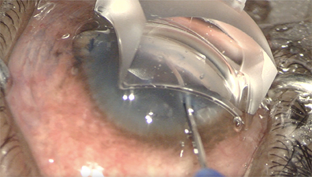
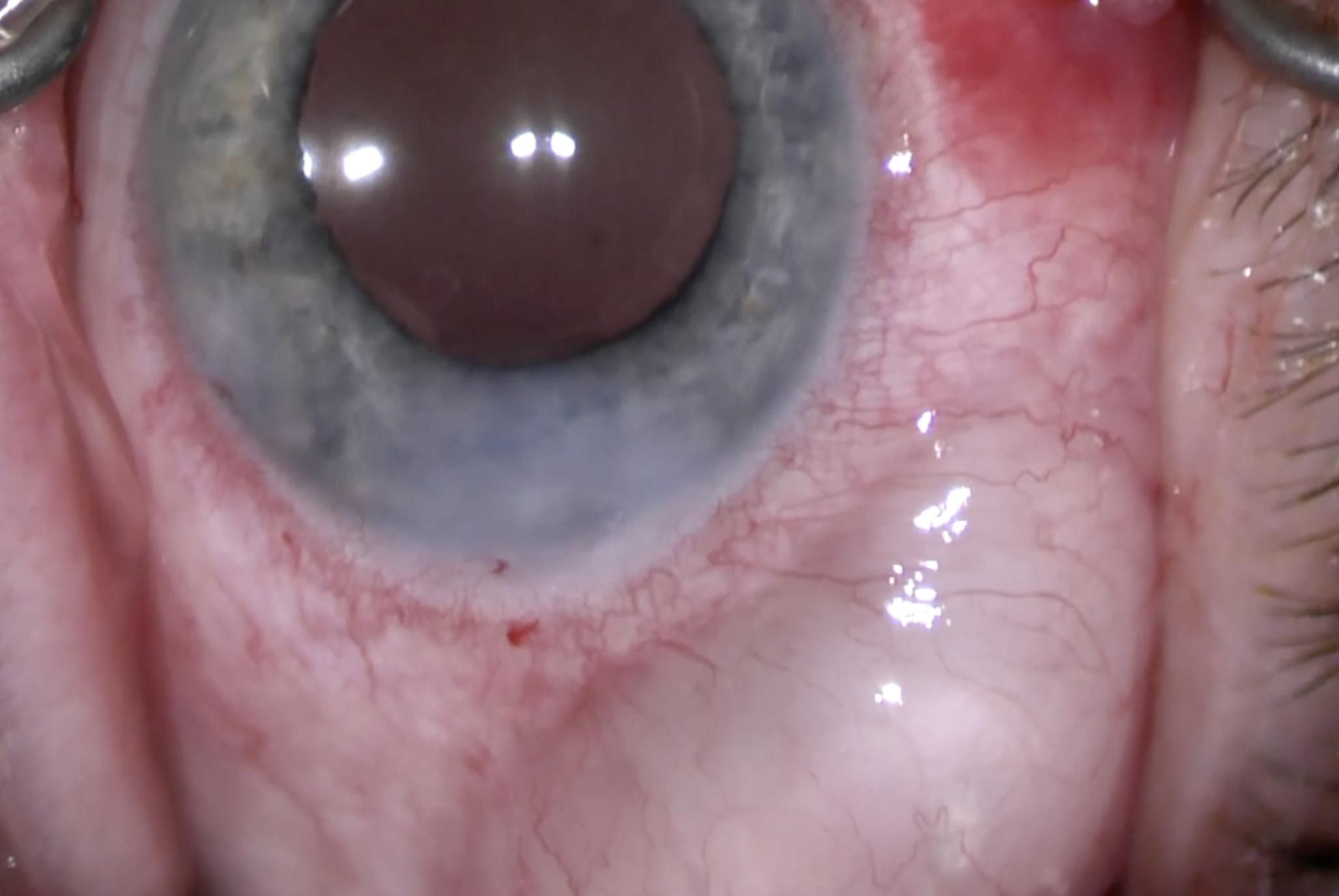
This is a 33-year-old male with congenital glaucoma who required shunt implantation to control his glaucoma. Cataract extraction was done to create room for in-sulcus tube placement due to high peripheral anterior synechiae (PAS).
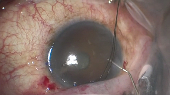
After careful measurement, the needle track was made. Minimal bleeding was observed at the tip of the needle and after needle withdraw. The tube was placed in the sulcus. No active bleeding was observed at the end of the operation.
At the first postoperative day, he developed hyphema and a blood clot was noted around the tube. After a week of conservative treatment, his clinical was not improved. His visual acuity was hand motion, and his IOP went up from 13 mmHg to 30 mmHg despite maximum medications.
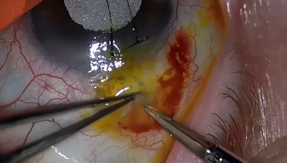
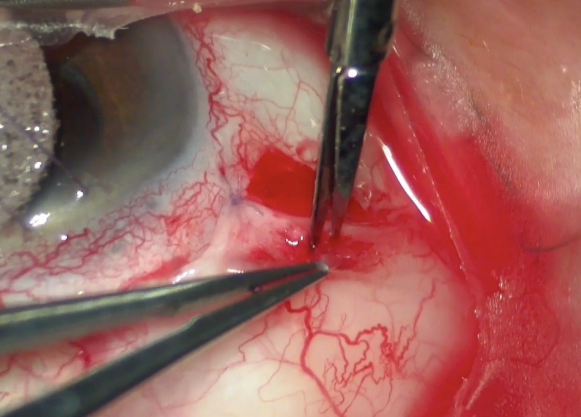
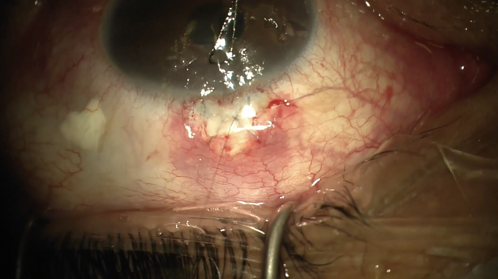
This prompted an anterior chamber irrigation in an attempt to remove the blood clot around the tube and in the anterior chamber. The blood clot was covered and stuck tight to the tube. Bleeding at the tube entry site is suspicious. It was confirmed by seeing blood oozing from around the tube after the clot was pulled out.
This eye is his better eye. I want to make sure that the bleeding will not occur again. ECP could be an answer, because not only can we directly visualize the bleeding site, but also ECP has a potential of being a hemostatic tool. This is attributed to the laser’s vascular coagulative effect, shown in this publication,1 that there was nonperfusion of the ciliary processes on endoscopic fluorescein angiography after treatment with ECP.
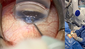
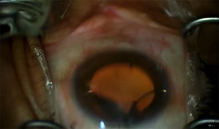
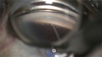
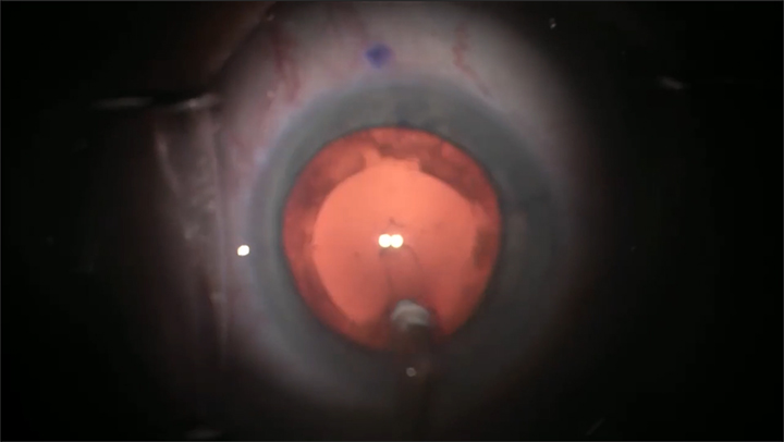
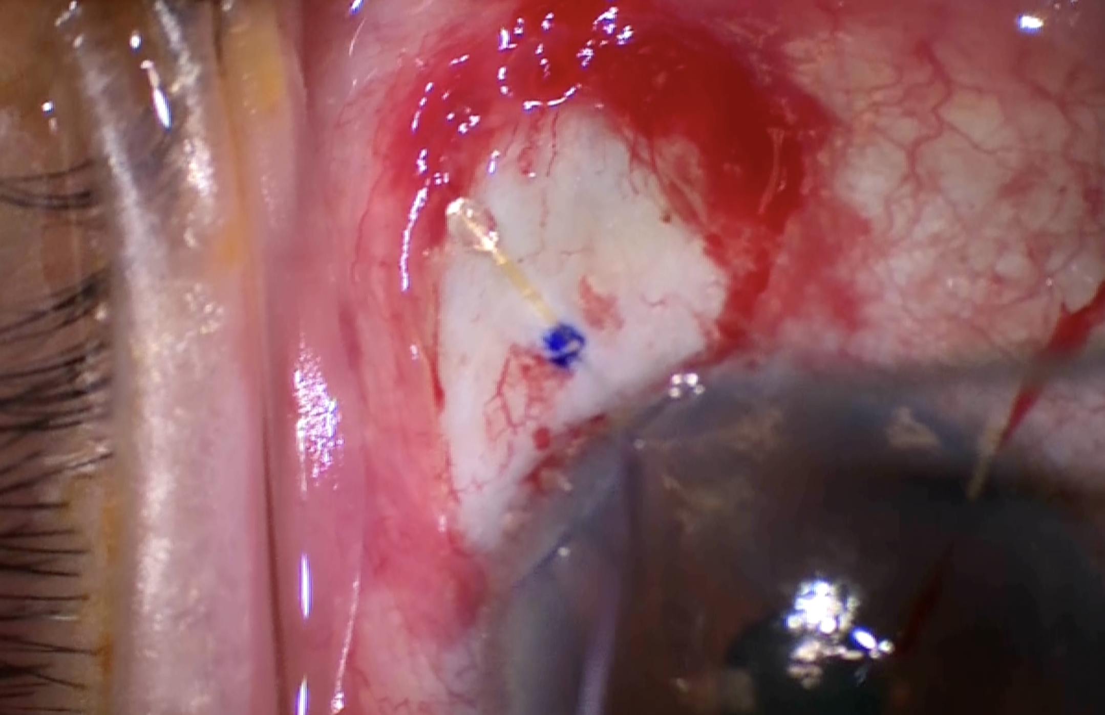
After deepening the sulcus with ophthalmic viscosurgical device (OVD), the ECP probe was placed to visualize the area of suspicious bleeding. ECP revealed a blood clot at the adjacent ciliary processes. Targeted photocoagulation achieved tissue whitening and shrinkage of the ciliary processes, with complete hemostasis.
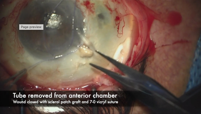
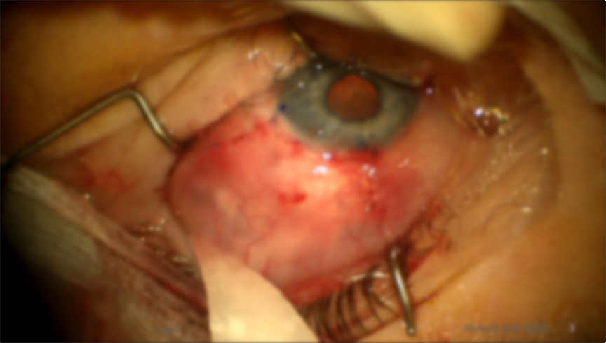
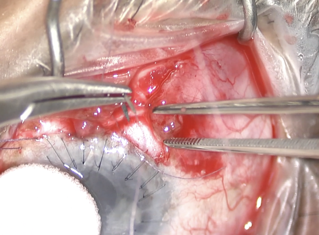
After OVD was removed, the eye became soft. No bleeding was observed. The wound was closed to ensure it was completely watertight. From postoperative day 1 until 4 months, there was no recurrent hyphema.
This shows that ECP can be an option to stop bleeding from ciliary processes. Thank you very much for watching. Please contact me if you have any questions or comments. GP

Reference
1. Lin SC, Chen MJ, Lin MS, Howes E, Stamper RL. Vascular effects on ciliary tissue from endoscopic versus trans-scleral cyclophotocoagulation. Br J Ophthalmol. 2006;90(4):496-500. doi:10.1136/bjo.2005.072777
Other Videos From Series